Home
/ Upper Leg Tendon Anatomy - Anatomy Of The Thigh Everything You Need To Know Dr Nabil Ebraheim Youtube - The four muscles all extend the lower leg.
Upper Leg Tendon Anatomy - Anatomy Of The Thigh Everything You Need To Know Dr Nabil Ebraheim Youtube - The four muscles all extend the lower leg.
Upper Leg Tendon Anatomy - Anatomy Of The Thigh Everything You Need To Know Dr Nabil Ebraheim Youtube - The four muscles all extend the lower leg.. Its muscle belly is on the back aspect of the upper arm. A visit to an orthopedic surgeon may be needed to accurately diagnose and treat your condition. They have a lot to do with how your hips move. Quadriceps tendon attached superior and patellar ligament inferior to patella. Notice the upper leg has a biceps muscle just like the upper arm does.
Your upper leg includes seven major muscles. A muscle strain (muscle pull or tear) is a common injury, particularly among people who participate in sports. The muscle is one of the four quadriceps muscles and is the largest muscle of that group. The thigh has three sets of strong muscles: The vastus laterails works with the other quad muscles to help extend your knee joint.
Tendinitis And Bursitis Treatment Cincinnati Tendinitis Dayton Oh from www.beaconortho.com The rectus femoris is located in the center of the thigh, while the vastus medialis is in the middle of the said body part. If you feel it you need to take care of the causes of this hard pain. Medial muscles adduct and rotate your thigh, and posterior flex your leg and extend your thigh. It is the junction of the thigh and the leg and is a hinge joint. 3d anatomy tutorial on the muscles of the thigh and the gluteal region from anatomyzone for more videos, 3d models and notes visit: On the medial edge of the posterior thigh is the gracilis muscle. Anatomy the four quadriceps muscles meet just above the kneecap (patella) to form the quadriceps tendon. The muscle is one of the four quadriceps muscles and is the largest muscle of that group.
The four muscles all extend the lower leg.
It serves to attach the plantaris, gastrocnemius (calf) and soleus muscles to the calcaneus (heel) bone. Upper leg anatomy and function. It also is active in maintaining thigh and kneecap position while walking and. A muscle strain or tear may cause your thigh to look deformed. 3d anatomy tutorial on the muscles of the thigh and the gluteal region from anatomyzone for more videos, 3d models and notes visit: It is the junction of the thigh and the leg and is a hinge joint. The rectus femoris is located in the center of the thigh, while the vastus medialis is in the middle of the said body part. Meanwhile, the vastus lateralis is on the side of the thigh, while the vastus intermedius is hidden below the rectus femoris(5). The hamstrings refer to 3 long posterior leg muscles, the biceps femoris, semitendinosus, and semimembranosus. Tendons are cords made of tough tissue, and they work as special connector pieces between bone and muscle. The four muscles all extend the lower leg. Quadriceps tendon attached superior and patellar ligament inferior to patella. Thigh pain that comes on suddenly and limits your ability to walk could be due to a pinched nerve in your back.
They consist of the rectus femoris, vastus intermedius, vastus lateralis and the vastus medialis. A muscle strain or tear may cause your thigh to look deformed. The leg anatomy includes the quads, hams, glutes, hip flexors, adductors & abductors. It is also visible on the medial edge of the thigh from the anterior. Your upper leg includes seven major muscles.
Top 8 Exercises To Build The Body Of A Greek God Leg Muscles Anatomy Muscle Anatomy Leg Anatomy from i.pinimg.com Thigh pain that comes on suddenly and limits your ability to walk could be due to a pinched nerve in your back. The four muscles all extend the lower leg. This important tendon in the back of the calf and ankle connects the plantaris, gastrocnemius, and soleus muscles to. Lateral (fibular) collateral ligament (fcl) upper part middle part lower part popliteus tendon (pt) upper part i. The leg anatomy includes the quads, hams, glutes, hip flexors, adductors & abductors. These muscles start at the bottom of your pelvis extending down the back of your thigh and along either side of your knee, to your lower leg bones. On the medial edge of the posterior thigh is the gracilis muscle. The thigh muscles don't just move your legs.
3d anatomy tutorial on the muscles of the thigh and the gluteal region from anatomyzone for more videos, 3d models and notes visit:
Your lower leg includes three main muscles, located behind your tibia or shinbone. Lateral (fibular) collateral ligament (fcl) upper part middle part lower part popliteus tendon (pt) upper part i. The quadriceps tendon attaches the quadriceps muscles to the patella. Upper leg anatomy and function. 3d anatomy tutorial on the muscles of the thigh and the gluteal region from anatomyzone for more videos, 3d models and notes visit: It's the area that runs from the hip to the knee in each leg. This mri wrist coronal cross sectional anatomy tool is absolutely free to use. The hamstring muscles in the back of the thigh, the quadriceps muscles in the front, and the adductor muscles on the inside. The hamstrings refer to 3 long posterior leg muscles, the biceps femoris, semitendinosus, and semimembranosus. The iliopsoas muscle flexes your hip, bends your trunk towards your thigh and rotates your thigh bone. The patella is attached to the shinbone (tibia) by the patellar tendon. A visit to an orthopedic surgeon may be needed to accurately diagnose and treat your condition. Upper leg anatomy and function the upper leg is often called the thigh.
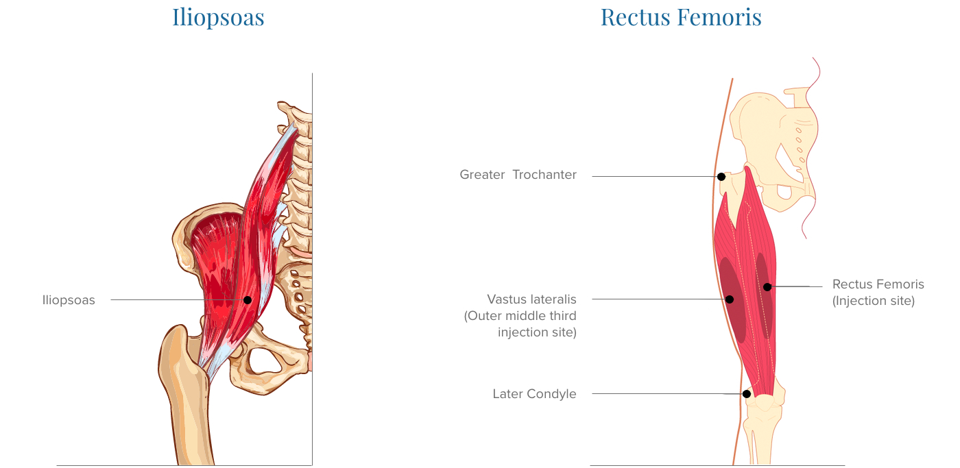
Tendons are cords made of tough tissue, and they work as special connector pieces between bone and muscle. In addition to these, the end of the iliopsoas muscle passes into the anterior. The vastus lateralis is a muscle located on the lateral, or outside, part of your thigh. Possibly the most important tendon in terms of mobility is the achilles tendon. Your upper leg includes seven major muscles.
The Anatomy Of The Lower Leg Muscles from www.verywellhealth.com Upper leg muscle pain is a very hard pain affect the leg pain as a whole. The hamstring muscles in the back of the thigh, the quadriceps muscles in the front, and the adductor muscles on the inside. Upper leg tendon anatomy : The iliopsoas muscle flexes your hip, bends your trunk towards your thigh and rotates your thigh bone. Tendons are thick bands of tissue that connect muscles to bone. The patella is attached to the shinbone (tibia) by the patellar tendon. They have a lot to do with how your hips move. •medial thigh muscles•adductor longus muscle•adductor magnus muscle•adductor.
The patella is attached to the shinbone (tibia) by the patellar tendon.
•medial thigh muscles•adductor longus muscle•adductor magnus muscle•adductor. The thigh muscles don't just move your legs. It is the junction of the thigh and the leg and is a hinge joint. It also is active in maintaining thigh and kneecap position while walking and. The four muscles all extend the lower leg. Anatomy the four quadriceps muscles meet just above the kneecap (patella) to form the quadriceps tendon. The hamstrings refer to 3 long posterior leg muscles, the biceps femoris, semitendinosus, and semimembranosus. This important tendon in the back of the calf and ankle connects the plantaris, gastrocnemius, and soleus muscles to. The iliopsoas muscle flexes your hip, bends your trunk towards your thigh and rotates your thigh bone. They consist of the rectus femoris, vastus intermedius, vastus lateralis and the vastus medialis. On the medial edge of the posterior thigh is the gracilis muscle. Tendons are cords made of tough tissue, and they work as special connector pieces between bone and muscle. Upper leg muscle pain is a very hard pain affect the leg pain as a whole.

/GettyImages-149678870-5702c33f5f9b581408a76e0b.jpg)